
Figure 1. HUVEC express von Willebrand factor but not Lex epitopes.
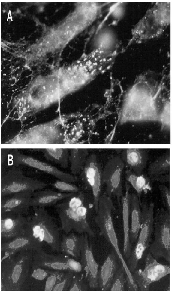
a) Immunofluorescence study of HUVEC endothelial cells immunostained with anti-von Willebrand factor polyclonal antibodies to confirm endothelial phenotype. Cells exhibit a homogeneous staining pattern localized in endothelial-specific organelles called Weibel-Palade bodies. Magnification: × 400. b) Immunofluorescence study performed on PMN adhered to HUVEC monolayers with mAb FC-2.15. A high level of Lex expression can be seen on PMN, while HUVEC did not stain. Magnification: × 200.