Figure 6.

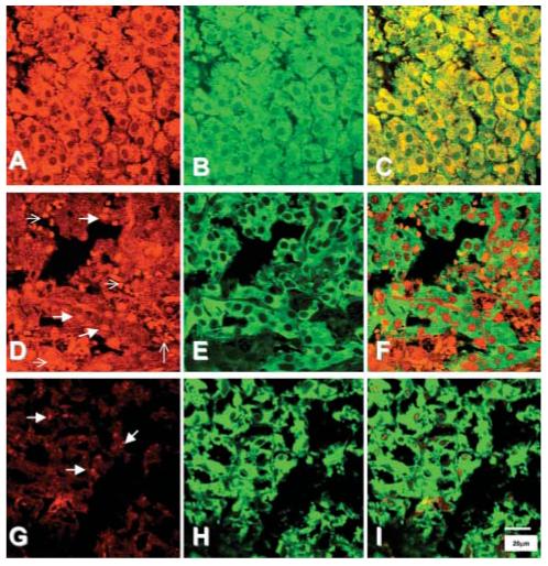
Immunohistochemical staining of the noradrenaline transporter (red, A, D, G), and tyrosine hydroxylase (TH) (green, B, E, H) in representative samples of the normal human adrenal medulla (A-C) and in phaeochromocytomas from patients with MEN 2 (D-F) and VHL syndrome (G-I). Staining of the transporter in the normal adrenal was present within the cytoplasm, and colocalized with staining for TH (C). In contrast, the pattern of staining for the noradrenaline transporter in tumour samples, of both MEN2 and VHL patients, was highly variable, and did not always appear to colocalize with TH. Thick arrows indicate granular staining for the noradrenaline transporter, thin arrows indicate red blood cells.
