
Figure 1.
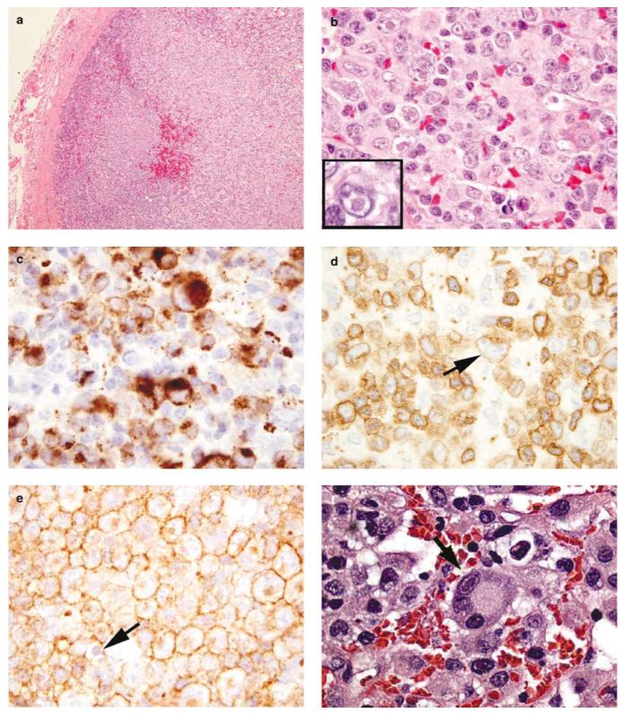
Histological and immunohistochemical findings. (a) and (b) HE-stained sections of the cervical lymph node from the Case no. 1 with markedly expanded paracortical areas containing a polymorphous population of atypical cells of varying sizes. Many of the cells contain large, eosinophilic, nuclear, and/or cytoplasmic inclusions, suggesting viral etiology. (c) Immunostaining for HHV-6 revels numerous positive atypical cells. Cytoplasmic inclusions are positive; however, antibody does not stain nuclear inclusions. (d) and (e). Immunohistochemical stains reveal that atypical cells, including cells with inclusions (arrows), are positive for CD3 (d) and CD4 (e) and negative for CD8 (not shown). (f) H&E-stained section of the liver biopsy from the Case No 2 demonstrating viral inclusions.