
FIGURE 1.
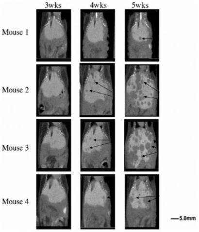
Serial DHOG hepatobiliary contrast-enhanced microCT scans reveal progression of MPC liver metastasis over time (arrows: tumors; Left: at 3 weeks, Middle: at 4 weeks, Right: at 5 weeks) Scale bar: 5.0 mm.
Official websites use .gov
A
.gov website belongs to an official
government organization in the United States.
Secure .gov websites use HTTPS
A lock (
) or https:// means you've safely
connected to the .gov website. Share sensitive
information only on official, secure websites.
Serial DHOG hepatobiliary contrast-enhanced microCT scans reveal progression of MPC liver metastasis over time (arrows: tumors; Left: at 3 weeks, Middle: at 4 weeks, Right: at 5 weeks) Scale bar: 5.0 mm.