
FIGURE 3.
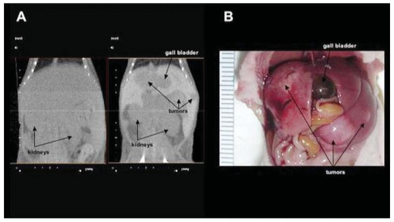
(a) MicroCT without and with DHOG contrast enhancement in the same mouse. Tumors are visualized only in the contrast-enhanced scan. (b) Macroscopic examination of the scanned mouse showing extensive liver metastases.
Official websites use .gov
A
.gov website belongs to an official
government organization in the United States.
Secure .gov websites use HTTPS
A lock (
) or https:// means you've safely
connected to the .gov website. Share sensitive
information only on official, secure websites.
(a) MicroCT without and with DHOG contrast enhancement in the same mouse. Tumors are visualized only in the contrast-enhanced scan. (b) Macroscopic examination of the scanned mouse showing extensive liver metastases.