
Figure 1. Membranous E-cadherin and F-actin localization in the morula and blastocyst stage embryos of hamsters.
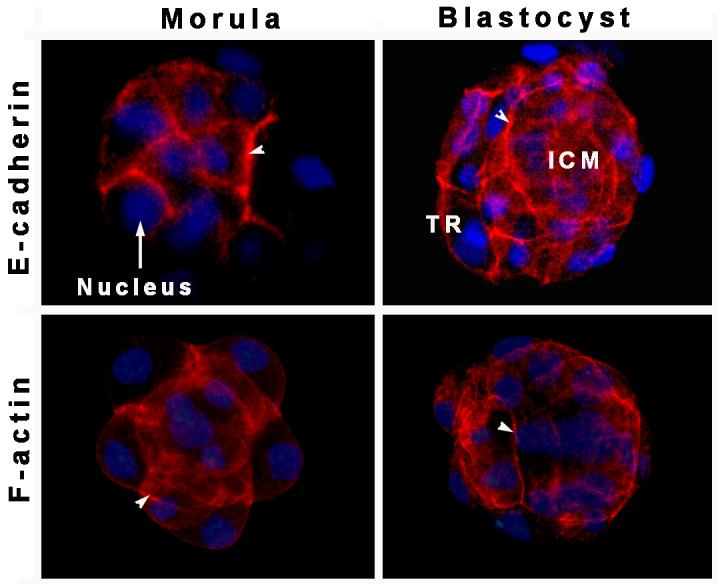
Arrowheads indicate E-cadherin (TRITC, red) and F-actin localization at cell-cell contact sites between blastomeres. A rat monoclonal E-cadherin antibody and Texas red-labeled phalloidin were used for E-cadherin and F-actin staining, respectively. Nuclei were stained with DAPI (blue). ICM, inner cell mass; TR, trophectoderm