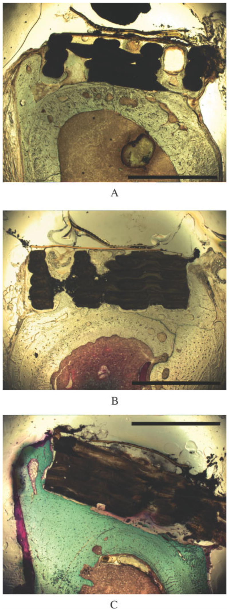
Figure 10.

Cross-sectional light micrographs of femora implanted with TGF-β1-enhanced scaffolds show bone formation. Plain PBT (a) and LC-PBT (b) scaffolds showed extensive bone growth into the pores and around the edges of the scaffolds, whereas VI-PBT scaffolds showed bone growth around the scaffold edge (c). The scale bar is 2 mm.
