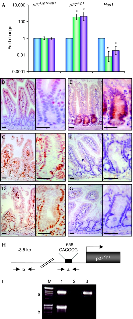
Figure 3.
Loss of Notch signalling in the crypt compartment results in loss of Hes1 expression and derepression of the cyclin-dependent kinase inhibitor p27Kip1. (A) Crypt regions of control (blue), N1N2 dKO (Notch1/Notch2lox/lox-vil-Cre-ERT2; green) and Rbpj KO (Rbpjlox/lox-vil- Cre-ERT2; purple) mice were laser microdissected and RNA isolated for quantitative reverse transcription–PCR. Relative levels of messenger RNA of p21Cip1/Waf1, p27Kip1 and Hes1 genes normalized with glyceraldehyde phosphate dehydrogenase are shown (logarithmic scale, *P<0.05; results represent the average of four independent experiments). (B–D) Immunohistochemical analysis of sections of the small intestine from (B) control, (C) N1N2 dKO and (D) Rbpj KO stained with an antibody against p27Kip1 bars. (E–G) Immunostaining for Hes1 on sections of the small intestine derived from (E) control, (F) N1N2 dKO and (G) Rbpj KO. The right-hand panels show a higher magnification of crypts from the left-hand panels. Scale bars, 50 μm. (H) Schematic representation of the p27Kip1 promoter indicating the primers used (a and b) for the chromatin immunoprecipitation (ChIP) analysis. The black rectangle corresponds to a class C Hes1-binding site (CACGCG; Murata et al, 2005). Primers indicated by ‘b', 3 kb upstream of the ATG, were used for control PCR. (I) ChIP assay. Epithelial cells from crypt enrichment preparations were processed for ChIP with an antibody against Hes1 (lane 3) and purified rabbit IgGs as a control (lane 2). PCRs of two distinct regions indicated by ‘a', containing the class C Hes1-binding site, and by ‘b', an Hes1-binding-site-free region, which was used as a negative control, are shown. Lane 1 corresponds to the input DNA.

