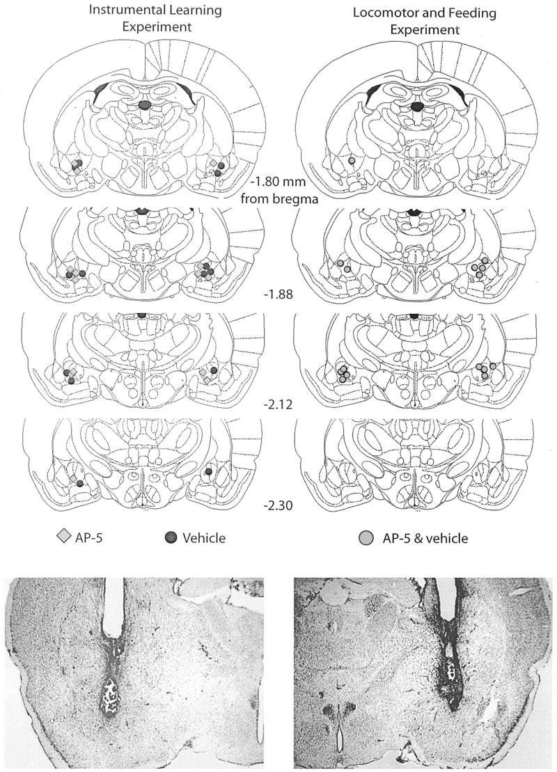
Figure 1.

Top: Histological reconstructions of cannula placements in the central nucleus of the amygdala (Experiment 1) are represented in schematic form. Histological sections were examined under a light microscope, and the site of the infusion was estimated. AP-5, n = 8; vehicle, n = 6; AP-5 + vehicle, n = 7. AP-5 = 2-amino-5 phosphonopentanoic acid. Reprinted from The Rat Brain in Stereotaxic Coordinates, 4th ed., G. Paxinos and C. Watson, Figures 26–29, Copyright 1998, with permission from Elsevier. Bottom: Photomicrograph examples of sites represented in the schematic diagrams.
