
Figure 1.
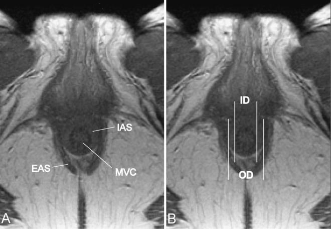
Axial MR scan. Pelvic magnetic resonance images (MRI) shown in the axial plane at the level of the sphincter complex. In all slices in which the internal anal sphincter (IAS) was clearly visible and could be distinguished from the mucosal vascular core (MVC) and the external anal sphincter (EAS) (A) measurements were taken (B). Thickness of the IAS was calculated as the difference between outer diameter of the IAS (OD) and inner diameter (ID).