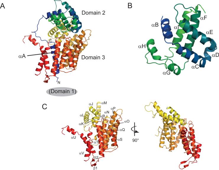
Figure 1. Structure of Cep3p.
A. Ribbon diagram of Cep3p, colored from blue at the N-terminus (54) to red at the C-terminus (608). Domain 1 (the N-terminal Zn2Cys6 cluster), absent in this construct, is located immediately before helix αA. B. Domain 2 (residues 78−229) has a structure similar to the globin fold. C. Domain 3 (residues 230−608) forms a left-handed solenoid composed of seven helical zig-zags which encircles αA. Disordered loops absent from electron density link αM to αN and αU to αV.

