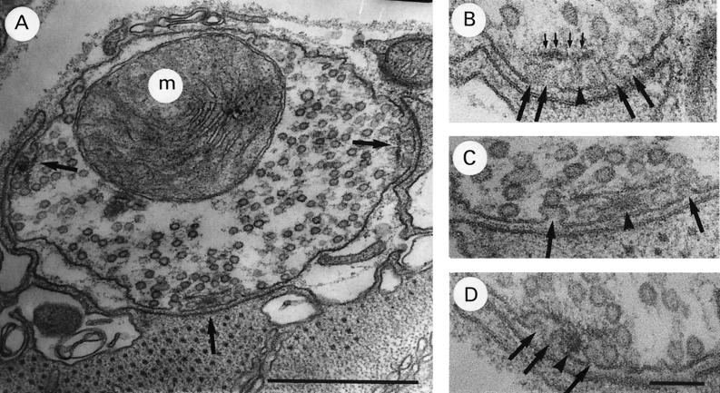
Figure 2.
Typical coxal terminal after 30 min exposure to 1 mM Ca2+/35 mM Mg2+ saline. (A) Terminal with three abnormal active zones (arrows) with invaginating plasma membrane under dense body plate. (B–D) Active zones in A shown at higher magnification. Note omega-shaped images (large arrows) under dense body plate (small arrows) Dense body base–arrowhead. [Bars: A, 0.5 μm (×68,000); and B–D, 100 nm (×135,000).]

