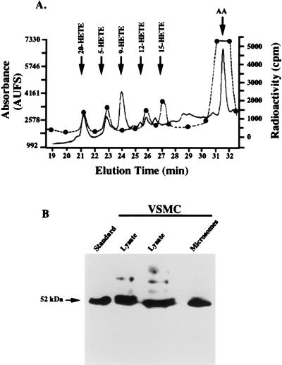
Figure 2.
Production of 20-HETE and expression of CYP450 in VSMCs. (A) Representative reverse-phase HPLC of products formed by VSMCs incubated with [14C]AA. Solid line, absorbance units full scale (AUFS); dashed line, radioactivity; arrow, position of authentic standards. (B) Detection of CYP450 4A in microsomes and lysates of VSMCs. Approximately 100–200 μg of proteins from microsomes and lysates were subjected to SDS/PAGE (10% gel) and detected by Western blotting using a rat CYP450 4A polyclonal antibody raised in goats. Lanes (from left to right) are clofibrate-treated liver microsomes as a standard, VSMC lysates (two lanes), and microsomes isolated from rabbit VSMCs.

