
Fig. 1.
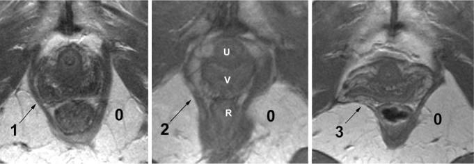
Examples of grade 1, 2, and 3 unilateral defects in axial images at the level of the midurethra. The urethra (U), vagina (V), and rectum (R) are labeled in the middlepanel of the figure. The blackarrow in each panel points to the left pubovisceral muscle in which there is loss of muscle bulk in comparison to the normal contralateral side