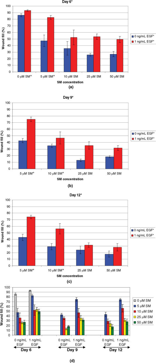
Figure 2.
Wound fill time-course study for NHEK cells exposed to various concentrations of SM and treated with 1 ng/mL of EGF. Cells were exposed to 0, 5, 10, 25, or 50 μM of SM, treated daily with 0 or 1 ng/mL of EGF, and stained with 0.1% crystal violet at 6 (a), 9 (b), or 12 (c) days after wounding. A significant difference* was observed between the 2 treatment groups, 0 and 1 ng/mL of EGF, on days 6, 9, and 12. However, no significant interactions were observed between SM concentrations and EGF doses, so it cannot be specifically stated that there was a significant difference between EGF doses at a particular SM concentration (ie, 0, 5, 10, 25, or 50 μM of SM). Significant differences between SM concentrations,† regardless of EGF dose, were observed at each day. (a) For day 6, 0 μM of SM† had significantly different wound fill than 5, 10, 25, and 50 μM of SM. In addition, 5 μM of SM† had significantly different wound fill than 10, 25, and 50 μM of SM. (b and c) For days 9 and 12, 5 μM of SM† had significantly different wound fill than 10, 25, and 50 μM of SM and 10 μM of SM† had significantly different wound fill than 25 and 50 μM of SM. Data points represent mean values ± SEM of 6 determinations from 2 separate experiments. A 2-factor ANOVA at each staining day was used to compare the SM concentrations and the 2 EGF doses. Statistical significance was defined as P ≤ .05 for all tests. (d) Summary graph of parts (a)–(c).

