
Figure 1.
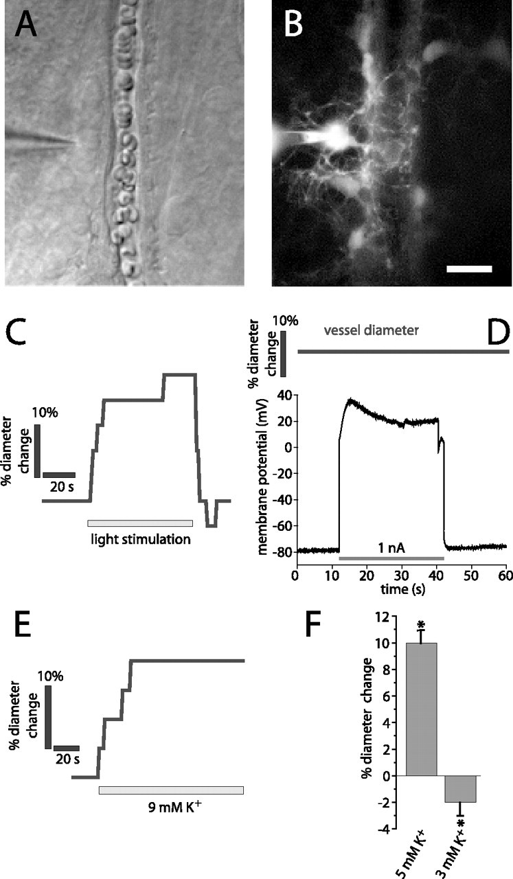
Light stimulation and K+ increase, but not glial cell depolarization, evokes vasodilation. A, B, Micrographs showing a whole-cell-patched astrocyte contacting an arteriole. The patch pipette is seen on the left. A, IR-DIC image. B, Fluorescence image showing the Lucifer-filled astrocyte contacting the arteriole. Additional astrocytes coupled to the patched cell are also seen. Scale bar: (in B) A, B, 10 μm. C–E, Experiments performed on the arteriole and astrocyte in A and B. C, Light stimulation evokes vasodilation. D, The astrocyte is depolarized by injection of 1 nA current. The arteriole diameter does not change during astrocyte depolarization. E, Ejection of 9 mm K+ in the region shown in A evokes vasodilation. F, Ejection of 5 mm K+ solution but not control solution (3 mm) evokes vasodilation in a series of experiments. Asterisks indicate significant change, p < 0.05. Error bars indicate SEM.