
Fig. 5.
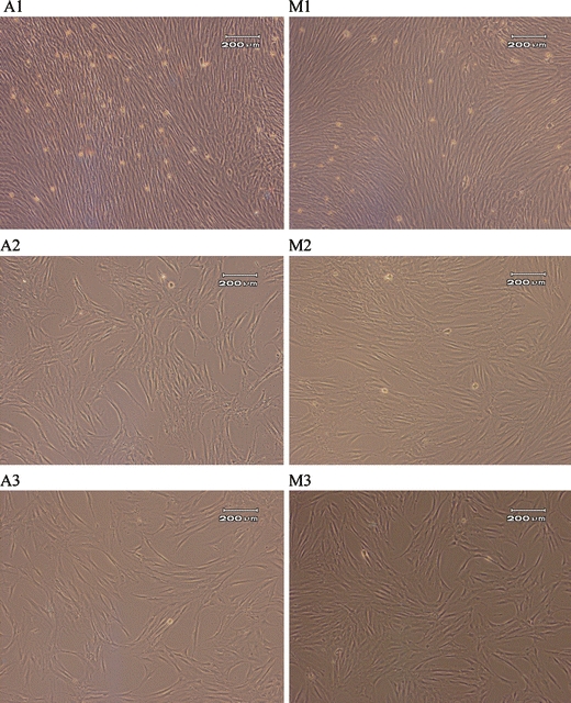
A representative selection of images of cells cultured using automated (A) and manual (M) culture protocols. 1: Closely associated cells in colony centres immediately prior to P1, 2: Cells in monolayer prior to P2, 3: Cells in monolayer prior to P3. Cells are morphologically similar at all points (sample 2)