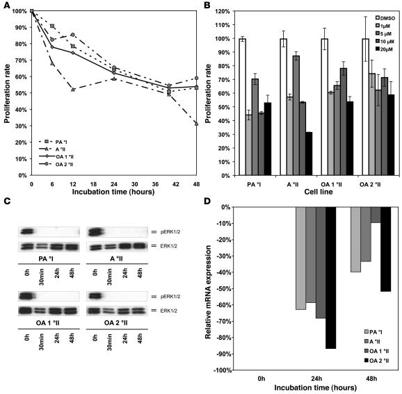
Figure 3. Pharmacological inhibition of MAPK signaling in cell lines derived from low-grade gliomas.
(A) Proliferation of glioma cells in vitro after treatment of 4 different cell lines derived from primary low-grade gliomas with the MEK1/2 inhibitor U0126 at a concentration of 20 μM as assessed by MTT assay over a time course of 48 hours. A °II, diffuse astrocytoma; OA, oligoastrocytoma. (B) Effective growth inhibition in all 4 cell lines can be achieved over a broad spectrum of concentrations ranging from 1 μM up to 20 μM. Medians and SDs of triplicate measurements at 48 hours are shown. (C) Dephosphorylation of ERK1/2 is readily detectable after 30 minutes and is maintained for 48 hours after a single dose of inhibitor at a concentration of 1 μM. (D) Maximal downregulation of CCND1 mRNA expression after treatment with MEK1/2 inhibitor U0126 at a concentration of μM was observed after 24 hours.

