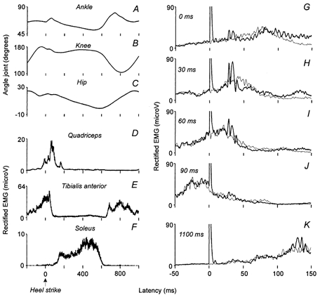
Figure 1. Recordings of the modulation of the angle joints, the ongoing EMGs and the CPN-induced facilitations in Q EMG activity during walking.

A–C, the variations of the angle of the ankle (A), of the knee (B) and of the hip (C) joints, expressed in degrees, are plotted against the latency (ms) after contact between the heel of the shoe and the treadmill (0 of the abscissa, Heel strike); the speed of the treadmill was set at 4 km h−1. D and E, the rectified EMG recordings (amplitude expressed in mV) of quadriceps (VL, D), tibialis anterior (TA, E) and soleus (Sol, F) muscles are also plotted against the latency (ms) after heel strike. G-K, the Q (VL) EMG activity conditioned by a CPN stimulation adjusted at 2.5 × MT (thick lines) and the control EMG (thin lines, average of 50 trials of each) are plotted against the latency (ms) after the stimuli (0 of the abscissa corresponds to the artefact of the stimulation).
