
Figure 2. Enhancement of presynaptic calcium current by intracellular CSP.
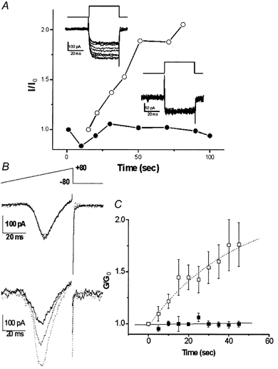
A, current traces from two calyces patched with pipettes containing either plain intracellular solution (inset lower right) or recombinant CSP (inset upper left) and recorded beginning immediately after seal rupture. The voltage protocol was a depolarizing step from −80 to 0 mV. Current amplitudes (I) were measured near the end of the step, normalized to that of the first trial (I0, trace with dark line) and were plotted (with CSP, ○; control, •). B, currents activated by a depolarizing ramp protocol (top panel) recorded in a control calyx (middle panel) or with CSP (lower panel). Traces were recorded immediately after seal rupture (t = 0, bold) and at 20 (dotted) and 45 s (fine-dotted) thereafter. C, a series of ramp depolarizations were given as in Fig. 2B at 5 s intervals to CSP-infused (□) and vector-control-infused (▪) calyx nerve terminals. The slope conductance, G, was determined as the slope of a straight line fitted to the ascending limb of the current peak between +35 and +40 mV and this was normalized to the value of G at t = 0 (G0). n = 4 for each treatment.