
Figure 4. Left (LV) and right ventricular (RV) stroke volumes (SVs) versus end-diastolic volume (EDV).
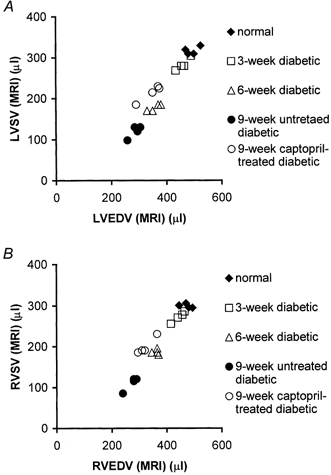
Comparative plots of MRI-measured left (A) and right (B) ventricular stroke volumes (SVs) and end-diastolic volumes (EDVs) of the five experimental groups. Data from rats diabetic from 7 and 10 weeks suggested impaired left and right ventricular diastolic and systolic function.