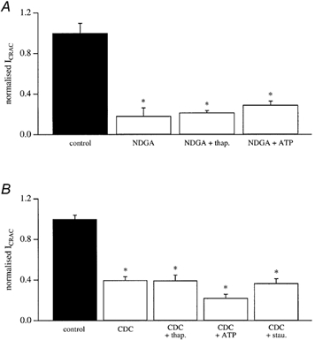
Figure 3. Lipo-oxygenase inhibition of ICRAC persists in the presence of thapsigargin, ATP and staurosporine.

A, the size of ICRAC in control cells, in those pre-exposed to 20 μm NDGA, and then after either 2 mm Mg-ATP or 2 μm thapsigargin have been included in the pipette solution following pre-exposure to NDGA. B, the size of ICRAC in control cells, after exposure to 20 μm CDC, and then after inclusion of either Mg-ATP or thapsigargin in the pipette solution following CDC pretreatment. Also included are data taken from cells first exposed to staurosporine and then CDC. Note that CDC still reduces ICRAC even in the presence of the kinase blocker. Each bar is the mean of 4–7 cells.
