
Figure 6. Changes in [Cl−]i on modifying basolateral Cl− concentration.
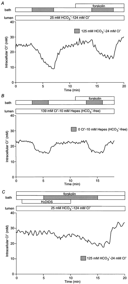
A, initially the bath and lumen of the duct segments were perfused with the standard HCO3−-buffered solution. The basolateral perfusate was then switched to the low Cl−-high HCO3− solution in the absence and presence of forskolin (1 μm). One of five experiments. B, with the bath and lumen initially perfused with the standard Hepes-buffered solution, the luminal perfusate was switched to the 0 Cl− Hepes-buffered solution in the absence and presence of forskolin (1 μm). One of four experiments. C, with the bath and lumen initially perfused with the HCO3−-buffered solution, the basolateral membrane was pretreated with H2DIDS (200 μm) and then the basolateral perfusate was switched to the low Cl−-Ihigh HCO3− solution in the presence of forskolin (1 μm). One of four experiments.