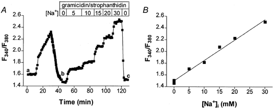
Figure 1. Traditional in vivo SBFI calibration.

A, representative experiment in a rabbit cell. At the end of a physiological protocol (region a-b) the cells were perfused with external solutions with various [Na+]o in the presence of 10 μm gramicidin and 100 μm strophanthidin (region b-c). The F340/F380 ratio increased with increasing [Na+]. B, linear fit of the calibration points in the same cell.
