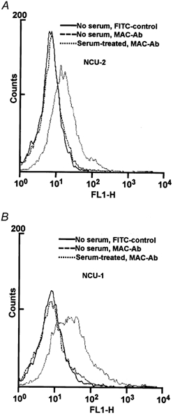
Figure 2. Flow cytometry of NCU-2 (A) and NCU-1 (B) cells after human serum treatment and incubation with anti-MAC antibody.

Both cell types harbour MAC in their membranes (dotted line). The fluorescence signal of cells incubated with anti-MAC antibody without serum treatment (dashed line) does not differ significantly from the signal of the FITC control (continuous line).
