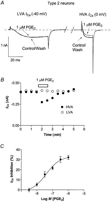
Figure 2. Calcium currents in Type 2 trigeminal neurons are inhibited by PGE2.

A, to elicit the low voltage-activated T-type ICa, neurons were stepped from −80 mV to −40 mV. Neurons recovered for 80 ms (approximately 65 ms has been removed for clarity) before stepping the membrane potential to 0 mV to elicit HVA ICa. T-type ICa was not inhibited by 1 μm PGE2. This trace was not leak subtracted and zero current is denoted by the dashed line.B, time course of inhibition of HVA ICa by 1 μm PGE2 in the same cell. C, Type 2 neurons were superfused with various concentrations of PGE2 for 1 min and then washed until the current returned to pre-drug treatment amplitude. Each point represents the mean and s.e.m. of 6–12 cells.
