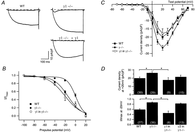
Figure 1. Expression of the γ1 subunit in γ1-deficient myotubes restores WT L-type Ca2+ current amplitude and steady-state inactivation.

A, representative current traces during a 400 ms depolarisation at −40 mV and at +20 mV from a WT and a γ1-deficient myotube, and from a γ1-deficient cell transfected with the γ1 subunit. The dashed line indicates zero. At −40 mV, a T-type Ca2+ channel is activated in the γ1-deficient cell. B, average steady-state inactivation (normalised to −100 mV prepulse potential) from WT (filled squares, n = 27), γ1-deficient cells (filled circles, n = 36) and from γ1-transfected γ1-deficient cells (open circles, n = 8). Data were fitted with a Boltzman equation. C, average I-V relationships of WT (filled squares, n = 30), γ1−/− (filled circles, n = 29) and γ1-transfected γ1-deficient (open circles, n = 9) myotubes. D, bar graphs showing the current density at +20 mV (top) and the steady-state inactivation after a prepulse of −20 mV (bottom) for WT and γ1-deficient myotubes, γ1-deficient myotubes expressing the γ1or the γ2 protein. Asterisks indicate statistical significance (P < 0.05), values in parentheses indicate number of cells.
