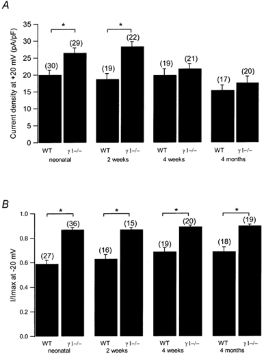
Figure 2. Age dependence of L-type Ca2+ current amplitude and steady-state inactivation.

A, current densities at +20 mV are plotted for WT and γ1-deficient myotubes from neonatal, 2-week-old, 4-week-old and 4-month-old mice. B, normalised steady-state inactivation after a 5 s prepulse to −20 mV for WT and γ1-deficient myotubes from neonatal, 2-week-old, 4-week-old and 4-month-old mice. Asterisks indicate statistical significance (P < 0.05), values in parentheses indicate number of cells.
