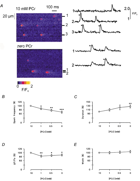
Figure 6. Effects of PCr on spontaneous Ca2+ sparks in permeabilized myocytes.

A, representative longitudinal line scan images of Fluo-3 fluorescence from the same myocyte in the presence of PCr (top) and in its absence (bottom). Selected plots of F/F0 (to the right of each image) were obtained by averaging over 3 pixels, centered on a Ca2+ release event. Comparison of the largest Ca2+ sparks obtained under both conditions (*) shows that sparks were typically smaller and more prolonged in the absence of PCr. B-E, accumulated data illustrating the effects of PCr on the frequency, duration, amplitude and width of spontaneous Ca2+ sparks. Error bars represent the means ± s.e.m., n = 8. *P < 0.05, **P < 0.02 and ***P < 0.005.
