
Figure 3. RNA and protein expression profile of KChIP2 isoforms.
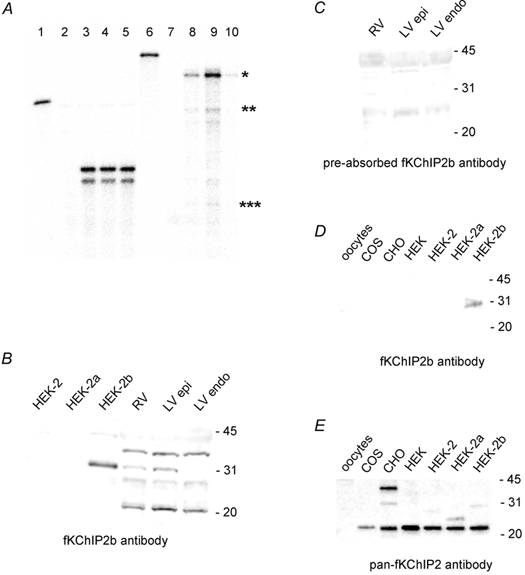
A, RNase protection assay for fKChIP2 isoforms across ferret ventricular walls. Lane 1: full-length undigested cyclophilin probe. Lane 2: hybridization of cyclophilin probe to yeast RNA. Lanes 3, 4, 5: hybridization of cyclophilin probe to 5 μg RV, LV epicardial and LV endocardial RNA, respectively. (Nucleotide mismatches between the ferret cyclophilin sequence and mouse primers used for cloning the cyclophilin probe can account for the double bands seen in the final assay.) Lane 6: full length undigested fKChIP2b probe. Probe was designed towards the first 247 nt of fKChIP2b and thus would hybridize to 247 nt of fKChIP2b, 169 nt of fKChIP2 and 73 nt of fKChIP2a. Lane 7: hybridization of fKChIP2b probe to yeast RNA. Lanes 8, 9, 10: hybridization of fKChIP2b probe to 10 μg RV, LV epicardial and LV endocardial RNA, respectively. The hybridization signals for fKChIP2b, 2, and 2a are indicated by one, two and three asterisks, respectively. For immunoblot analysis, 10 μg HEK 293, COS, CHO, or Xenopus oocyte or 75 μg RV, LV epicardium, or LV endocardium protein was separated in each lane. HEK-2, −2a, or −2b refers to HEK 293 cells transfected with fKChIP2, 2a or 2b. B, immunoblot with fKChIP2b antibody. C, immunoblot with fKChIP2b antibody pre-incubated with fKChIP2b peptide used for antibody production. Exposures were equal for B and C. D, immunoblot with fKChIP2b antibody on protein from uninjected oocytes; non-transfected COS, CHO, or HEK 293 cells; and transfected HEK 293 cells. Exposure based on detection of HEK-2b as positive control. E, immunoblot with pan-fKChIP2 antibody on protein from uninjected oocytes; non-transfected COS, CHO, or HEK 293 cells; and transfected HEK 293 cells. Exposure based on detection of fKChIPs in transfected HEK 293 cells. fKChIP2, 2a and 2b are 29, 26, and 31 kDa, respectively.