
Figure 5. Large preceding depolarization does not affect the extent of ATP-induced ImVDCC potentiation and inhibition.
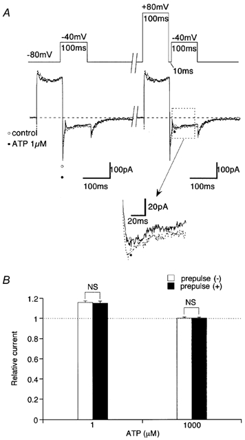
A, voltage protocol (upper trace) and corresponding current traces before (○ and continuous curve) and after (• and dotted curve) addition of 1 μm ATP. Inset indicated by arrow is magnification from a part boxed by dotted line. B, relative amplitude change of ImVDCC after addition of 1 μm (left) or 1 mm (right) ATP with (□) or without (▪) a 100 ms prepulse to 80 mV. Experiments carried out with Na+ as charge carrier (divalent cation-free conditions) in the presence of 10 μm nifedipine in the bath. NS, no statistically significant difference with unpaired t test. n = 4.