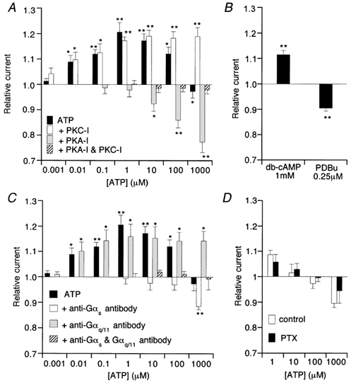
Figure 6. Effects of protein kinase inhibitors (A) activators (B), G-protein antibodies (C) and overnight pertussis toxin treatment (D) on ATP-induced modulation of ImVDCC.

Recording conditions were the same as in Fig. 4. PKC-I, protein kinase C inhibitory peptide (1 μm); PKA-I, protein kinase A inhibitory peptide (41.3 nm); PTX, pertussis toxin. In C, antibodies against Gαs and Gαq/11 were diluted 1:35 in the pipette solution. In D, mesenteric arteriolar myocytes were incubated with (PTX) or without (control) pertussis toxin (500 ng ml−1) at 10°C for 24h. *P > 0.05 and **P < 0.01 with t test for paired data (n = 5) before and after application of a given concentration of ATP, dbcAMP or PDBu.
