
Figure 1. Experimental design and injection sites.
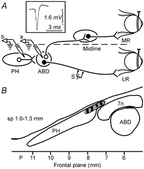
A, schematic representation of the animal preparation. Teflon-coated stainless-steel coils were fixed to the scleral margin of both eyes. A stimulating electrode (S) was implanted on the VIth nerve to retrogradely stimulate motoneurons in the abducens nucleus (ABD). A glass micropipette was introduced in the abducens nucleus (a) where its position was confirmed by the antidromic field potential recording (inset). After withdrawal, the pipette was introduced in the prepositus hypoglossi nucleus (PH, b) for drug injection; LR, lateral rectus muscle; MR, medial rectus muscle. B, schematic mapping of the injection sites in the rostral PH. Six representative injection sites are shown. A total of 17 drug injections were performed within the hatched area, in a saggital plane (sp) between 1.0 and 1.3 mm from midline and a frontal plane between 7 and 8 mm posterior to bregma. 7n, facial nerve.