
Figure 7. Co-localization of cyclic GMP and glutamic acid decarboxylase in neurons in the medial vestibular nucleus projecting to the ipsilateral prepositus hypoglossi nucleus.
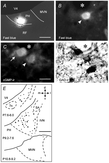
A, coronal section showing the Fast Blue injection area in the PH nucleus of one cat. B-D, photomicrographs showing a neuron (arrowhead) triple labelled with Fast Blue, cyclic GMP (cGMP-ir) and glutamic acid decarboxylase (GAD-ir) in the medial vestibular nucleus (MVN) ipsilateral to the injection side. A neuron positive for GAD-ir and cGMP-ir (filled arrow) and another one positive only for GAD-ir (open arrow) are also shown. The asterisk indicates a blood vessel. E, distribution of the neurons triple labelled with Fast Blue, cGMP-ir and GAD-ir in the MVN ipsilateral to the PH nucleus injected with Fast Blue. Each section contains the neurons found in the indicated frontal plane intervals. Data correspond to cats 36 (triangles) and 37 (dots). Calibration bars: A, 500 μm; B-D, 25 μm; E, 1 mm. 7n, facial nerve; d, dorsal; IVN, inferior vestibular nucleus; l, lateral; m, medial; RF, reticular formation; SA, stria acustica; v, ventral; V4, fourth ventricle.