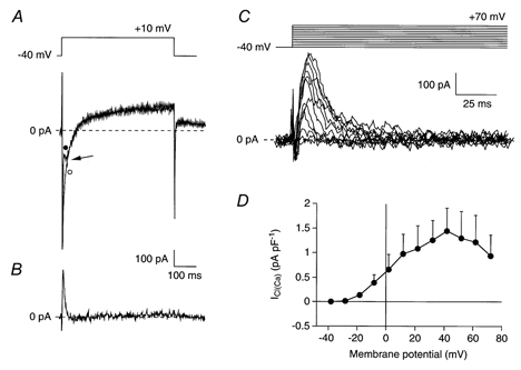
Figure 2. Presence of the Ca2+-activated Cl− current (ICl(Ca)) in SA node cells.

A, superimposed current traces of an SA node cell (Cm, 110 pF) elicited by voltage steps from −40 to +10 mV in absence (•) and presence (○) of 0.2 mm DIDS. Arrow indicates an outwardly directed transient current, which is superimposed on the inward peak current. This transient current is blocked by DIDS. B, DIDS-sensitive ICl(Ca) current obtained by digital subtraction of the two current traces of panel A. C, ICl(Ca) measured as the DIDS-sensitive current at potentials between −40 and +70 mV in the same cell as shown in panels A and B. The current activates around −20 mV and the peak amplitude increases upon further depolarization to +40 mV. At more depolarized potentials, the peak amplitude of ICl(Ca) decreases. D, average I-V relationship of ICl(Ca) in 7 cells.
