
Figure 7. Schematic model.
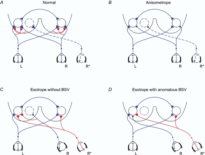
Proposed neural projections to account for present results, showing image projection into each eye and the neural projection, omitting the lateral geniculate nucleus, to the binocular integrating unit represented by the circles in the left and right visual cortices. Excitatory projections are shown in blue and inhibitory projections in red, without any implication as to how the inhibition is mediated. Magnitude of input to visual cortex is represented qualitatively by the size of the projection terminal. Binocular integrating centres in the two hemispheres are shown to be connected through the corpus callosum. L, left eye; R, right eye; R* is right eye with deviation of image onto nasal retina (A and B) or fovea (C and D). A, normal subject showing balanced left and right eye excitatory inputs which are attenuated by the action of high threshold mutual inhibition. The displaced image in the nasal retina of the right eye gives rise to a projection to the left visual cortex. This is shown not to be connected to the binocular integrating centre driven by the foveae of the left and right eyes. B, anisometrope amblyope showing reduced excitatory inputs both from the non-amblyopic eye and amblyopic eye and an absence of mutual inhibition to account for the enhanced binocular facilitation. As in A, the nasally directed image in the right eye has no adverse effect on contrast sensitivities. C, esotropic amblyope without BSV showing reduced excitatory inputs both from the non-amblyopic eye and amblyopic eye. In this case, there is a powerful inhibitory action of the esotropic eye input on the binocular integrating centre. Both binocular integrating centres are subjected to inhibition from foveal stimulation of the right eye. A similar scheme would apply to exotropes without BSV except that there is stimulation of the temporal retina and the binocular interactions are reflected onto reverse cortices. D, esotropic amblyope with anomalous BSV showing reduced excitatory inputs both from the non-amblyopic eye and amblyopic eye but this time leading to binocular facilitation due to establishment of new retinal correspondence; however, both binocular integrating centres are subjected to inhibition from foveal stimulation of the right eye as in C.