
Figure 4. Measurement of IPSC reversal potential.
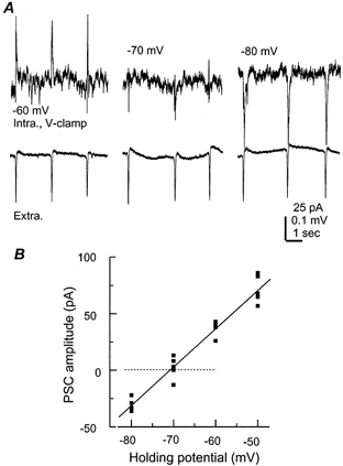
A, periodic IPSCs (top) and nearby rhythmic field potentials (bottom) simultaneously collected from a 21-day-old hippocampal isolate. The pyramidal neuron was recorded using the perforated (gramicidine) method and voltage clamped at different potentials as indicated. B, the amplitudes of IPSCs were measured and then plotted vs. the corresponding holding potentials. The line through data points was a linear regression fit (r = 0.97).