
Figure 10. Regionally originated rhythmic field potentials.
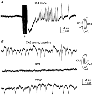
Hippocampal tissues were prepared from two mice aged 21 days old. A, extracellular record was collected from a CA1 tissue strip, showing no spontaneous- but evoked-field responses following repetitive stimulation of local circuitry (arrow, 20 Hz, 1 s). The recording setting and the longitudinal cut were schematically illustrated (right). B, extracellular records collected from a CA3 tissue strip before, following perfusion of bicuculline methiodide (BMI, 10 μm, 5 min) and after washing out bicuculline.