
Figure 1. Coincidence detection in the chick and the chick embryo.
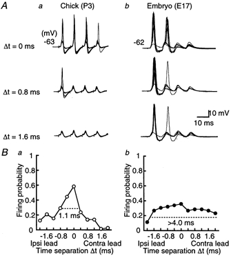
A, ten superimposed voltage traces of action potentials of the NL neuron generated by a pair of stimuli applied on both ipsilateral and contralateral projections from NM. Time differences (Δt) between the two sides of stimulation are indicated for the chick (a) and the embryo (b). Trains of four stimuli were applied repeatedly at 3 s intervals. Firing probability from the contra- and the ipsilateral stimuli alone was 0.13 (40 stimuli) and 0.18 (40 stimuli), respectively for the chick, and 0.06 (16 stimuli) and 0.06 (16 stimuli) for the embryo. Here and in subsequent figures, resting membrane potential is indicated to the left of each panel. B, firing probabilities as a function of Δt were calculated from the neurons in A. Note that the firing probability peaked at around Δt = 0 in both age groups, but had a more pointed peak in the chick (a) than in the embryo (b). The response windows are indicated by horizontal dashed lines. Ipsi, ipsilateral; Contra, contralateral.