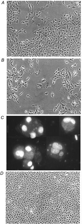
Figure 2. Effect of deprivation of compatible osmolytes on the morphology of cells exposed to hypertonicity.

A, cells were seeded and cultured for 24 h in isotonic (0.3 osmol (kg H2O)−1) medium and then incubated for a further 72 h in either (B) depleted hypertonic (0.5 osmol (kg H2O)−1) medium or (D) depleted hypertonic medium with added betaine and myo-inositol (each at 0.1 mmol l−1). The cells were then photographed under phase contrast. Cells that had detached after 72 h of hypertonic incubation in the absence of compatible osmolytes (C) were treated with the fluorescent DNA stain Hoechst 33342. Magnification: A, B and D, × 100; C, × 400. All the cells in field C exhibit the typical chromatin condensation of apoptosis.
