
Figure 3. Distribution of Kit-positive cells in C57BL/6 mouse antrum at the greater curvature, midway between greater and lesser curvature and at the lesser curvature.
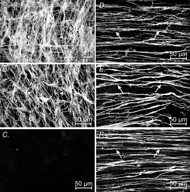
Kit-positive ICC at the level of the myenteric plexus at the greater curvature (A), midway between the greater and lesser curvature (B) and at the lesser curvature (C). Note the decrease in the distribution of ICC-MY (arrowheads) from the greater until they were not detectable at the lesser curvature. Distribution of ICC-IM (arrows) within the circular muscle layer at the greater curvature (D), midway between the greater and lesser curvature (E) and at the lesser curvature (F). A and D, B and E and C and F are confocal digital composites at the same site in the stomach. A, B and C are composites of 5 × 1 μm steps through the myenteric plexus region and D, E and F are composites of 15 × 1 μm steps within the circular muscle layer. Scale bar = 50 μm in all images.