
Figure 3. Outward IK1 transients observed using perforated-patch recording.
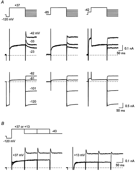
A, pre-pulse dependence of the transient outward currents observed during repolarizing voltage steps applied subsequent to a depolarizing pulse (+37 mV, 200 ms). A hyperpolarizing (−120 mV) or a depolarizing (−42 mV) pre-pulse was applied in the left or the right column, respectively. Currents recorded using repolarizing steps at voltages between −23 and −42 mV are superimposed in the middle row, and those at below −62 mV are superimposed in the lower row. B, dependence of the amplitude of the transient outward current on the duration of the depolarizing pulse (left panel, +37 mV, right panel, +13 mV). Currents recorded using depolarizing pulses of different durations (100, 200 and 300 ms) are superimposed in each panel. Currents shown in this figure were recorded from the same cell placed in an external solution containing nicardipine, chromanol 293B and E4031. The holding potential was set at the resting potential (−85 mV in this cell). Inward currents observed during hyperpolarizing pre-pulses are truncated in the middle row of A and B.