
Figure 3. The dual effect of 5-MT was mimicked by PKA activators and blocked by PKA inhibition.
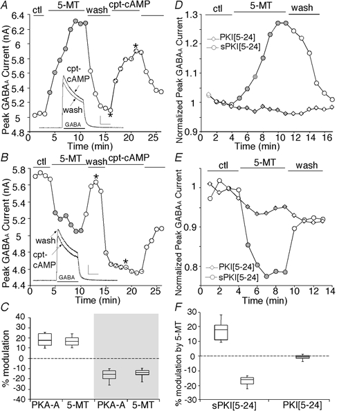
A and B, plot of peak GABAA current as a function of time and drug application. The 5-HT4 agonist 5-MT (20 μm) reversibly enhanced (A) or reduced (B) GABAA currents in the PFC neuron. Following recovery, application of the membrane-permeant cAMP analogue cpt-cAMP (200 μm) produced an effect that was similar to 5-MT, enhancing (A) or reducing (B) GABAA currents. Inset: representative current traces taken from the records used to construct A or B (at time points denoted by *). Scale: 1 nA, 1 s C, box plot summary of the percentage modulation of GABAA currents by PKA activators (PKA-A) or 5-MT. Note that these PKA activators (cpt-cAMP or sp-cAMPS) mimicked both the enhancement (n = 7) and reduction (n = 10) of GABAA currents caused by 5-MT. D and E, plot of peak GABAA current as a function of time and drug application in neurons dialysed with PKI[5–24] or sPKI[5–24]. The specific PKA inhibitory peptide PKI[5–24] (20 μm), but not the scrambled control peptide sPKI[5–24] (20 μm), eliminated 5-MT-induced enhancement (D) or reduction (E) of GABAA currents. F, box plot summary of the percentage modulation of GABAA currents by 5-MT in 5-HT4 mRNA-positive neurons dialysed with PKI[5–24] (n = 10) or sPKI[5–24] (enhancement: n = 5; reduction: n = 6).