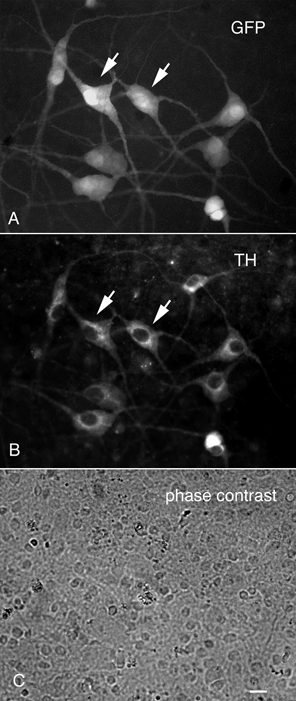
Figure 4. LC neurons express GFP and tyrosine hydroxylase in culture.

The LC and surrounding brain were dispersed and cultured on glass coverslips. A, LC neurons retained their GFP expression. B, immunostaining for tyrosine hydroxylase (TH) showed that most neurons showing TH immunoreactivity also showed GFP expression. C, a phase contrast micrograph of the area shown in A and B reveals a large percentage of unlabelled cells, many of them glial cells. Scale bar: 5 μm.
