
Figure 1. Group I activation causes a reduction of visual responses.
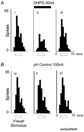
A, iontophoretic application of DHPG (50 nA) reversibly inhibited visual responses. The histograms show cumulative counts of action potential spikes (200 ms epochs) in response to 10 visual stimuli of a neurone under control conditions (i), during DHPG iontophoresis (ii) and recovery from DHPG effects (iii). Visual stimuli were presented during the period marked by the bar, and were 5 by 10 deg bars moving at 22.5 deg s−1 for 4 s every 10 s. The moving bars were of preferred orientation and direction. B, application of H+-Na+ from an acidified saline solution did not affect visual responses of the same neurone as in A.