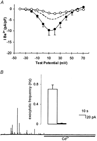
Figure 4. H2O2 mimics hypoxic and amyloid peptide augmentation of current density, but does not induce Cd2+-resistant evoked exocytosis.

A, mean current density vs. voltage plots (with vertical s.e.m. bars, where visible behind symbols) obtained from seven cells exposed to 40 μM H2O2 for 24 h. Recordings were made in the absence (•) or presence (○) of 2 μM nifedipine. Dashed line indicates, for ease of comparison, control current density (taken from Fig. 3A). B, amperometric recording of exocytosis from a representative PC12 cell exposed to 40 μM H2O2 for 24 h. Secretion was evoked by exposure to a perfusate containing 50 mm K+ (application period commencing at the beginning of the trace), and 200 μM Cd2+ was applied for the period indicated by the horizontal bar. Inset shows mean ± s.e.m. exocytotic frequency in H2O2-treated cells before (□) and during (▪) exposure to Cd2+.
