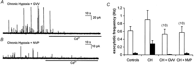
Figure 5. Inhibitors of γ secretase prevent hypoxic augmentation of exocytosis.

A, amperometric recording of exocytosis from a representative PC12 cell which had been cultured under chronically hypoxic conditions but in the additional presence of the γ secretase inhibitor, GVV (10 μM). Secretion was evoked by exposure to a perfusate containing 50 mm K+ (application period commencing at the beginning of the trace). B, as A, but the cell was incubated with another γ secretase inhibitor, NVP (10 μM). In both traces cells were exposed to 200 μM Cd2+ in the continued presence of 50 mm K+ for the period indicated by the horizontal bars. C, bar graph showing mean (with vertical s.e.m. bars, taken from the number of cells indicated above each bar) exocytotic frequency in the two cell groups indicated in A and B before (□) and during (▪) exposure to Cd2+. Control and CH data taken from Fig. 1 for ease of comparison.
