
Figure 7. Polarized expression of AQP4 in rat brain.
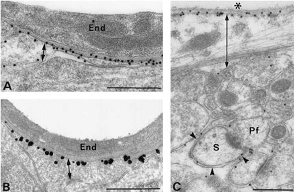
A and B, anti-AQP4-immunogold electron microscopy demonstrates AQP4 in glial membranes facing blood vessels but not in membranes facing neuropil. C, anti-AQP4-immunogold electron microscopy demonstrates AQP4 in sub-pial astrocyte membranes. Scale bars: A and B, 0.5 μm; C, 1 μm. Reprinted from Journal of Neuroscience with permission (Nielsen et al. 1997b).