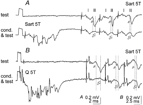
Figure 4. Temporal facilitation of disynaptic components of group II intermediate zone field potentials.

A, records of the field potential illustrated in Fig. 3A, conditioned by three stimuli at a conditioning-testing interval of 17 ms. B, another intermediate zone field potential, at a conditioning-testing interval of 25 ms; recorded at a more ventral location (depth 3.1 mm, probably lamina VIII) where synaptic actions of group I afferents no longer preceded synaptic actions of group II afferents (Edgley & Jankowska, 1987a). Note that in both cases the monosynaptic components of field potentials, which were originally evoked by group II afferents (small arrows) were abolished by the conditioning stimuli. Note also that the delayed components (large arrows) were larger after the second and third stimuli than after the first stimulus. Other indications are as in Fig. 3.
