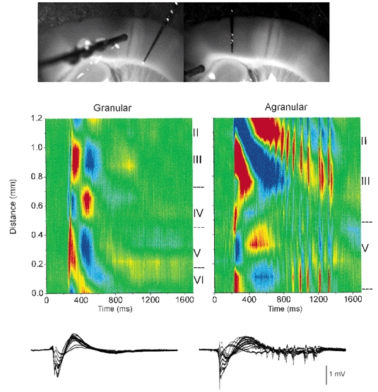
Figure 6.

CSD analysis of spontaneous discharges recorded during disinhibition in granular and agranular regions of the same slice
Upper panel, photomicrographs of the slice preparation taken during the experiment showing the location of the 16-channel silicon probe used to derive the CSD analyses shown below. A lesion separated the granular and agranular regions. A stimulating electrode (shown in left panel) served to demonstrate the effectiveness of the lesion to eliminate communication between both regions by stimulating in one region and recording in the other. Middle panel, CSD analyses of spontaneous events recorded in the granular (left) and agranular (right) region of the same slice during BMI+CGP35348. Lower panel, the field potential recordings used to derive the CSD are overlaid. Current sinks are in red, sources are blue and zero is green.
